
Introduction: How to Perform X-ray Examinations
X-ray examinations are an essential diagnostic tool in modern medicine. They provide detailed images of the internal structures of the body, allowing doctors to identify and diagnose a wide range of medical conditions. X-ray examinations are commonly used to diagnose bone fractures, lung infections, and dental problems, among many other conditions.
What is an X-ray Examination?
An X-ray examination, also known as radiography, is a medical imaging technique that uses X-rays to produce images of the inside of the body. X-rays are a type of electromagnetic radiation that can penetrate through the body, creating images of the internal structures such as bones and organs.
Why are X-ray Examinations Important?
X-ray examinations are important because they allow doctors to diagnose medical conditions that may not be visible from the outside. They are non-invasive and relatively quick, making them a useful tool for diagnosing a wide range of conditions. X-ray examinations can also be used to monitor the progress of medical treatments and to ensure that conditions are healing properly.
Performing X-ray examinations requires specialized training and equipment. In this step-by-step guide, we will walk you through the process of conducting X-ray imaging, from preparing the patient to interpreting the results.
Preparing for the X-ray Examination
Before undergoing an x-ray examination, there are certain things you should expect and prepare for. X-ray imaging is a safe and painless procedure that uses low doses of radiation to produce images of the inside of your body. Here’s what you need to know before going for an x-ray examination:
What to Expect Before the X-ray Examination
Prior to the x-ray examination, you will be asked to fill out a medical history questionnaire. This is important as it helps the radiologist determine if you have any medical conditions or are taking any medications that may affect the imaging results. You will also be asked to remove any jewelry or metal objects from your body as they can interfere with the imaging process.
It is important to inform your doctor if you are pregnant or suspect you may be pregnant as radiation can be harmful to a developing fetus. In such cases, your doctor may recommend alternative imaging methods such as ultrasound or MRI.
How to Prepare for the X-ray Examination
Preparing for an x-ray examination is simple and straightforward. Here are some tips to help you prepare:
- Clothing: Wear loose, comfortable clothing that can be easily removed. Avoid clothing with zippers, buttons, or any metal fastenings as they can interfere with the imaging process.
- Diet: You can eat and drink normally before the examination unless your doctor advises otherwise. However, if you are having an abdominal x-ray, you may be asked to avoid eating or drinking for a few hours before the exam.
- Medications: Inform your doctor if you are taking any medications, especially those containing iodine or have had iodine contrast material injected for a previous imaging procedure. Depending on the type of x-ray examination, your doctor may ask you to stop taking certain medications temporarily.
It is important to follow all instructions given by your doctor or radiologist before the x-ray examination. Failure to do so may result in inaccurate imaging results, which can affect your diagnosis and treatment.

Performing the X-ray Examination
As a professional radiographer, performing an x-ray examination requires a well-coordinated approach to ensure accurate imaging results. The process involves three main steps: positioning the patient, adjusting the x-ray machine, and capturing the x-ray image.
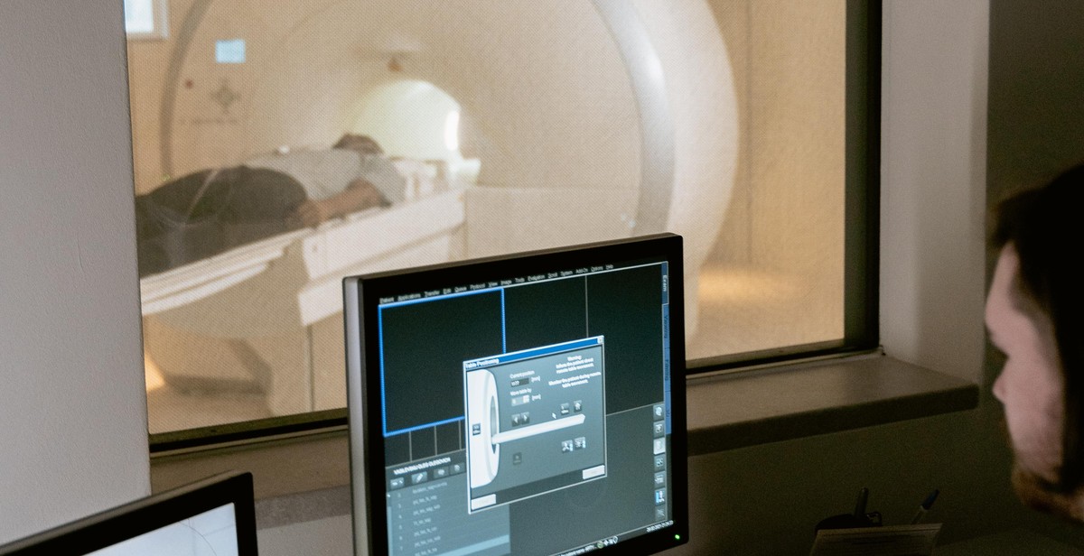
Step 1: Positioning the Patient
Proper patient positioning is crucial to obtaining clear and accurate x-ray images. The radiographer should communicate effectively with the patient, explaining the procedure and ensuring they are comfortable and relaxed. The patient should be positioned correctly, according to the area of the body being examined, to prevent any distortion or misalignment of the image.
For example, when imaging the chest, the patient should stand with their back against the x-ray plate and their arms raised above their head. When imaging the spine, the patient should lie on their back with their legs straight and their arms at their sides. Proper patient positioning not only improves the quality of the image, but also reduces exposure to unnecessary radiation.
Step 2: Adjusting the X-ray Machine
After positioning the patient, the radiographer should adjust the x-ray machine to the appropriate settings. The machine should be set to the correct kilovoltage (kV) and milliamperage (mA) based on the area of the body being imaged and the patient’s body size. The radiographer should also ensure the collimator is adjusted to the correct size to avoid unnecessary radiation exposure to surrounding tissues.
Moreover, the radiographer should ensure that the x-ray machine is functioning properly and that all safety precautions are in place. The patient should be given a lead apron to wear to protect their reproductive organs from unnecessary radiation exposure.
Step 3: Capturing the X-ray Image
Once the patient is positioned correctly and the x-ray machine is adjusted, the radiographer can capture the x-ray image. The patient should remain still and hold their breath for a few seconds to avoid any motion blur on the image. The radiographer should also ensure that the x-ray beam is centered on the area of interest to obtain a clear image.
After capturing the image, the radiographer should review it to ensure it is of high quality and meets the diagnostic requirements. If the image is not clear or if there are any technical issues, the radiographer may need to repeat the procedure.
Conclusion
Performing an x-ray examination requires a coordinated approach from the radiographer to ensure accurate imaging results. Proper patient positioning, adjusting the x-ray machine, and capturing the x-ray image are the three main steps involved in the process. By following these steps, radiographers can obtain high-quality images that are crucial for accurate diagnosis and treatment.
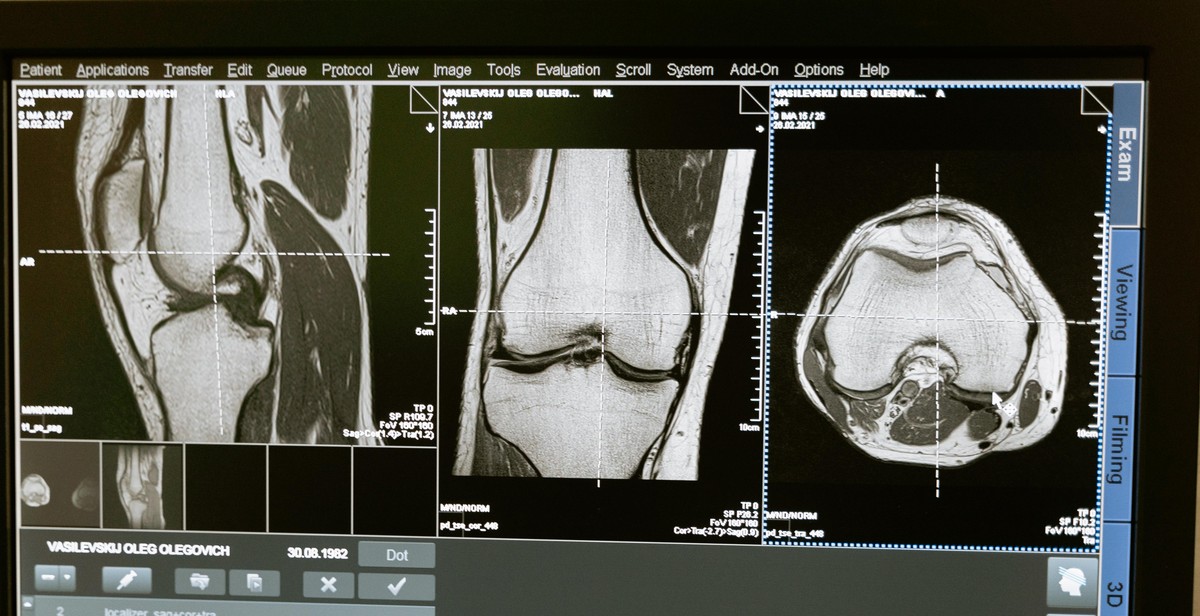
Interpreting the X-ray Image
Interpreting an X-ray image can be challenging, especially for someone who is not familiar with the anatomy of the body part being examined. However, with practice and experience, interpreting an X-ray image becomes easier. Here are a few things to look for when interpreting an X-ray image:
What to Look for in an X-ray Image
- Bones: Bones show up as white on an X-ray image. Look for any breaks, fractures, or dislocations. Also, check for any signs of arthritis or bone tumors.
- Tissues: Soft tissues, such as muscles and organs, show up as shades of gray on an X-ray image. Look for any abnormalities, such as swelling or fluid buildup.
- Foreign Objects: X-ray images can also detect foreign objects in the body, such as bullets or glass shards.
Identifying Abnormalities in the X-ray Image
When interpreting an X-ray image, look for any abnormalities that may indicate a medical condition. Here are a few examples:
| Abnormality | Description |
|---|---|
| Fracture | A break in the bone that appears as a discontinuity or gap in the white line of the bone. |
| Dislocation | A bone that is out of its normal position. This may be indicated by an asymmetry in the joint space. |
| Arthritis | A condition that causes inflammation and stiffness in the joints. This may be indicated by a loss of joint space or bone spurs. |
| Tumor | An abnormal growth of tissue that may appear as a mass or abnormal shadow on the X-ray image. |
| Pneumonia | An infection of the lungs that may appear as areas of white or gray on the X-ray image. |
It’s important to note that not all abnormalities will be visible on an X-ray image. Some conditions, such as early-stage cancer or a blood clot, may not show up on an X-ray image and may require additional testing.
Overall, interpreting an X-ray image requires a trained eye and an understanding of anatomy and medical conditions. If you have any concerns about an X-ray image, be sure to consult with a qualified healthcare professional.
Conclusion
Performing X-ray examinations requires precision, attention to detail, and a thorough understanding of the equipment and procedures involved. By following the step-by-step guide outlined in this article, you can ensure that your X-ray imaging is conducted safely and accurately, yielding high-quality results that can aid in the diagnosis and treatment of a wide range of medical conditions.
Remember to always prioritize safety when working with X-ray equipment, including the use of protective gear and proper handling of potentially hazardous materials. Additionally, it is important to stay up-to-date with the latest advances in X-ray technology and techniques, as this field is constantly evolving and improving.
Key Takeaways
- Proper preparation, including patient positioning and equipment calibration, is crucial for successful X-ray imaging.
- Effective communication with patients and other medical professionals can help ensure that X-ray procedures are conducted efficiently and accurately.
- Continued education and training are essential for staying up-to-date with the latest advances in X-ray technology and techniques.
Remember to prioritize safety, precision, and communication when performing X-ray examinations, and always strive for the highest quality results possible.
| X-ray machine | X-ray technician | X-ray film |