How to Interpret X-Ray Images: Understanding Radiographic Anatomy
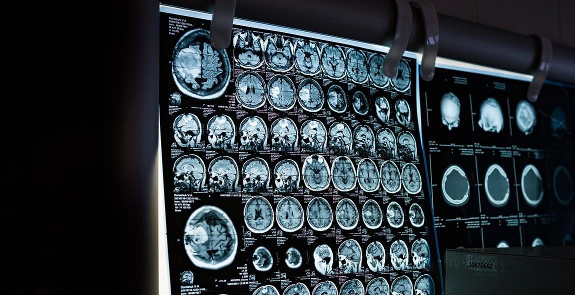
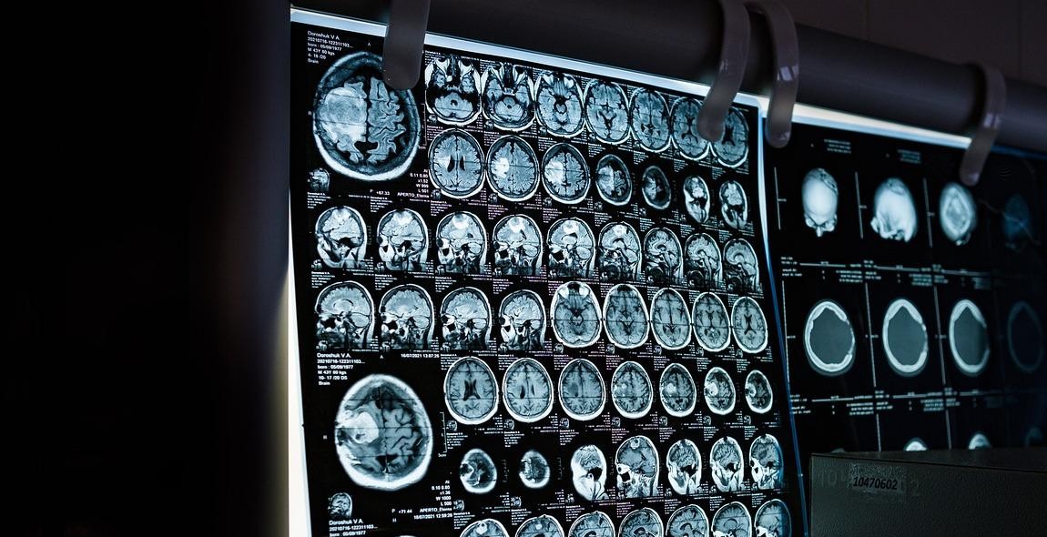
X-ray imaging is a crucial diagnostic tool used in medical settings to detect and diagnose various medical conditions. However, interpreting x-ray images can be daunting, especially for individuals without a medical background. Understanding radiographic anatomy is essential in interpreting x-ray images accurately.
The Importance of Radiographic Anatomy
Radiographic anatomy involves the study of the internal structures of the body as viewed through x-ray imaging. It is critical to understand the basic anatomy of the body and how it appears on x-ray images to interpret the results correctly.
Having a basic understanding of radiographic anatomy helps healthcare professionals to identify abnormalities in the patient’s internal structures correctly. It also helps to ensure that the images are taken correctly, minimizing the need for repeat imaging and reducing the patient’s exposure to radiation.
Interpreting X-Ray Images
Interpreting x-ray images requires a trained eye and knowledge of radiographic anatomy. The process involves analyzing the images for any abnormalities or changes in the internal structures of the body.
It is essential to note that x-ray images are not always conclusive, and further testing may be required to make a definitive diagnosis. However, a basic understanding of radiographic anatomy can help healthcare professionals to identify potential issues and determine the next steps in the diagnostic process.
This article aims to provide insights into radiographic anatomy and how it relates to interpreting x-ray images accurately.
What is Radiographic Anatomy?
Radiographic anatomy is the study of anatomical structures using X-ray images. It involves the interpretation of X-ray images to identify and locate different structures within the body. Radiographic anatomy is an essential part of medical imaging, and it plays a critical role in the diagnosis and treatment of diseases.
Definition of Radiographic Anatomy
Radiographic anatomy is the study of the internal structures of the body using X-ray images. It involves the use of X-rays to create images of the body, which can be used to identify and locate different structures within the body. Radiographic anatomy is a branch of anatomy that deals with the study of the body’s internal structures, including bones, organs, and tissues.
Importance of Radiographic Anatomy
Radiographic anatomy is an essential tool in medical imaging, and it plays a critical role in the diagnosis and treatment of diseases. X-ray images can provide valuable information about the body’s internal structures, which can help doctors to identify and diagnose various medical conditions. Radiographic anatomy is particularly useful in identifying bone fractures, tumors, and other abnormalities in the body.
In addition to its diagnostic value, radiographic anatomy is also used in medical research to study the structure and function of different organs and tissues in the body. It is an important tool in the development of new medical treatments and therapies.
| Benefits of Radiographic Anatomy |
|---|
| Helps in the diagnosis and treatment of diseases |
| Provides valuable information about the body’s internal structures |
| Useful in identifying bone fractures, tumors, and other abnormalities |
| Important tool in medical research and development |
Overall, radiographic anatomy is an essential tool in modern medicine, and it has revolutionized the way we diagnose and treat diseases. With its ability to provide detailed images of the body’s internal structures, radiographic anatomy has become an indispensable tool for doctors, researchers, and medical professionals around the world.

How to Interpret X-Ray Images: Understanding Radiographic Anatomy
Interpreting X-ray images can be a challenging task, especially for individuals who lack medical training. However, with a basic understanding of radiographic anatomy, identifying normal structures and abnormalities in the images becomes much easier. In this section, we will discuss the key steps involved in interpreting X-ray images.
Understanding Radiographic Anatomy
Before interpreting X-ray images, it is crucial to have a basic understanding of radiographic anatomy. Radiographic anatomy refers to the appearance of different structures in the body when viewed through an X-ray. Some of the key structures that can be seen in X-ray images include bones, soft tissue, and air-filled spaces.
Bones appear as white structures on X-ray images, while soft tissue appears as shades of gray. Air-filled spaces, such as the lungs, appear as black structures. Understanding the appearance of these structures in X-ray images is essential for identifying abnormalities.
Identifying Normal Structures
Identifying normal structures is the first step in interpreting X-ray images. This involves examining the image for any abnormalities and comparing it to a standard image of a healthy individual. Some of the key structures that need to be examined include bones, joints, and soft tissue.
When examining bones, it is important to look for any signs of fractures, dislocations, or abnormal growth patterns. Joints should be examined for any signs of swelling or inflammation, while soft tissue should be examined for any signs of fluid accumulation or masses.
Identifying Abnormalities
After identifying normal structures, the next step is to look for abnormalities. Abnormalities can be classified as either congenital or acquired. Congenital abnormalities are present at birth and may be caused by genetic factors, while acquired abnormalities develop over time and may be caused by injury or disease.
Some of the common abnormalities that can be identified in X-ray images include fractures, tumors, infections, and arthritis. Fractures appear as breaks in bones, while tumors appear as abnormal growths. Infections can be identified by the presence of inflammation or fluid accumulation, while arthritis can be identified by changes in the joint space.
| Abnormality | Appearance on X-ray |
|---|---|
| Fractures | Breaks in bones |
| Tumors | Abnormal growths |
| Infections | Inflammation or fluid accumulation |
| Arthritis | Changes in joint space |
In conclusion, interpreting X-ray images requires a basic understanding of radiographic anatomy, identification of normal structures, and identification of abnormalities. By following these key steps, healthcare professionals can accurately diagnose and treat various medical conditions.

Common X-Ray Examinations and their Interpretations
Interpreting X-rays is a crucial part of radiography. X-rays are used to diagnose a wide range of medical conditions, and they are often the first line of defense when it comes to detecting abnormalities in the body. Here are some of the most common types of X-ray examinations and their interpretations:
Chest X-Ray
A chest X-ray is a common diagnostic tool that is used to assess the health of the lungs, heart, and other structures in the chest. This type of X-ray can help detect conditions such as pneumonia, lung cancer, and heart failure. When interpreting a chest X-ray, a radiologist will look for abnormalities in the lungs, such as fluid or air-filled spaces. They will also assess the size and shape of the heart and look for signs of heart disease.
Abdominal X-Ray
An abdominal X-ray is used to assess the digestive system and other structures in the abdomen. This type of X-ray can help detect conditions such as bowel obstruction, kidney stones, and abdominal tumors. When interpreting an abdominal X-ray, a radiologist will look for abnormalities in the organs and structures of the abdomen, such as the liver, spleen, and kidneys. They will also assess the size and shape of the digestive system, including the stomach, small intestine, and large intestine.
Bone X-Ray
A bone X-ray is used to assess the health of the bones and joints. This type of X-ray can help detect conditions such as fractures, arthritis, and bone tumors. When interpreting a bone X-ray, a radiologist will look for abnormalities in the bones and joints, such as breaks, cracks, or signs of wear and tear. They will also assess the alignment of the bones and look for signs of joint damage or inflammation.
Overall, X-rays are an essential tool in the diagnosis and treatment of many medical conditions. If you have any concerns about your health, it is important to speak with your healthcare provider about the appropriate diagnostic tests for your specific needs.
Conclusion
Interpreting X-ray images can be a daunting task, especially for those who are new to the field. However, with practice and a good understanding of radiographic anatomy, anyone can become proficient in interpreting X-ray images.
It is important to remember that X-ray images are just one part of the diagnostic process, and they should always be interpreted in conjunction with a patient’s medical history and physical examination.
When interpreting X-ray images, it is essential to pay close attention to the position of the patient, the quality of the image, and any abnormalities or anomalies that may be present. Familiarizing yourself with the anatomy of the body and the various structures that can be seen on X-ray images is also crucial.
Finally, it is essential to stay up-to-date with the latest advancements in radiography and to continue learning and expanding your knowledge base. This will not only improve your ability to interpret X-ray images but will also help you provide the best possible care to your patients.
- Always interpret X-ray images in conjunction with a patient’s medical history and physical examination.
- Pay close attention to the position of the patient, the quality of the image, and any abnormalities or anomalies that may be present.
- Stay up-to-date with the latest advancements in radiography and continue learning and expanding your knowledge base.
By following these guidelines, you can become a skilled and proficient interpreter of X-ray images, providing your patients with the best possible care.
