How to Create Stunning Radiology Artwork: Combining Science and Aesthetics
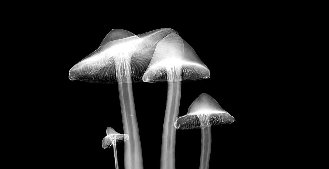
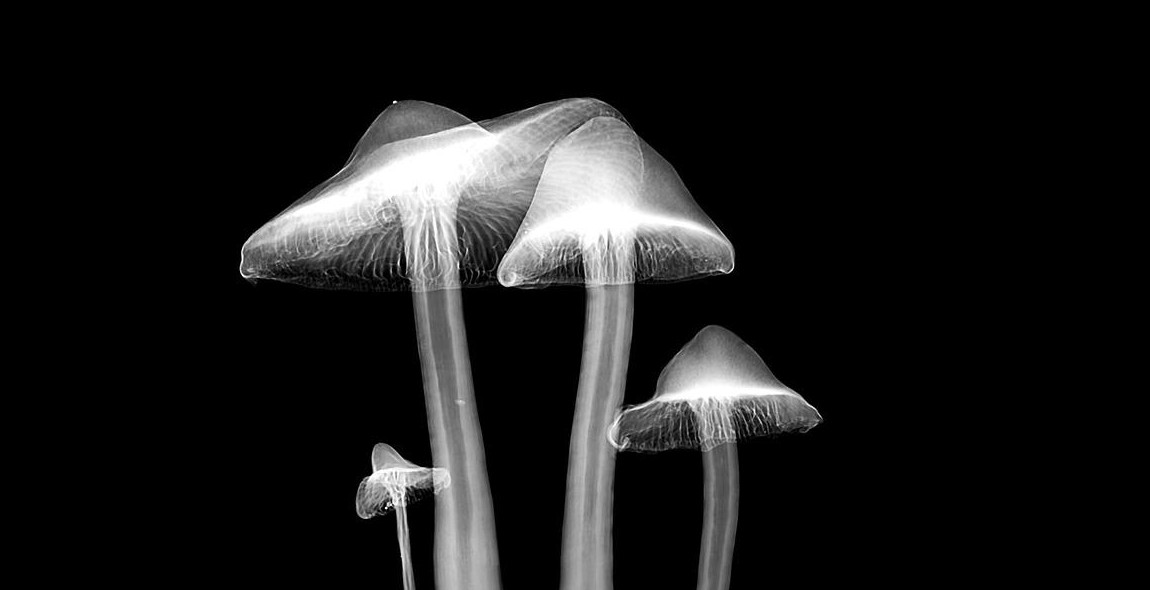
As a professional article writer and content creator, I have had the opportunity to explore the fascinating world of radiology artwork. Radiology is the branch of medicine that uses imaging techniques such as X-rays, CT scans, and MRIs to diagnose and treat diseases. Radiology artwork, on the other hand, is the creative application of these imaging techniques to produce stunning and visually appealing artwork.
Combining science and aesthetics, radiology artwork is a unique and innovative way to showcase the beauty of the human body and its inner workings. From the intricate patterns of the human brain to the delicate structures of the lungs, radiology artwork can transform medical images into captivating works of art.
The Importance of Radiology Artwork
Radiology artwork serves not only as a visual representation of the human body but also as a tool for education and communication. By transforming complex medical images into easy-to-understand visual representations, radiology artwork can help healthcare professionals and patients alike to better understand and communicate about medical conditions and treatments.
In this article, I will share my personal experience and expertise in creating stunning radiology artwork. From choosing the right images to selecting the best printing techniques, I will provide a comprehensive guide to help you create your own beautiful radiology artwork.
Understanding Radiology Artwork
Radiology is a medical specialty that focuses on the use of medical imaging to diagnose and treat diseases. It involves the use of various imaging technologies such as X-rays, CT scans, MRI scans, and ultrasound to visualize the internal structures of the body.
What is Radiology Artwork?
Radiology artwork is a unique form of art that combines the science of radiology with the aesthetics of art. It involves creating stunning and visually appealing images from medical imaging data. Radiology artwork is created using specialized software that allows radiologists and artists to manipulate and enhance medical images to create beautiful and detailed images.
Radiology artwork can take many forms, including posters, prints, and digital images. It can be used for educational purposes, as well as for decoration in medical facilities and offices. Radiology artwork is also used to raise awareness about diseases and medical conditions and to promote research and development in the field of radiology.
Why is Radiology Artwork Important?
Radiology artwork is important because it helps to bridge the gap between science and art. It allows people to appreciate the beauty and complexity of the human body in a way that is both informative and visually appealing. Radiology artwork can also help to demystify medical imaging and make it more accessible to the general public.
Furthermore, radiology artwork has important applications in medical education and research. It can be used to teach medical students and healthcare professionals about the human body and various diseases and conditions. Radiology artwork can also be used in research to visualize and analyze medical data in new and innovative ways.
In summary, radiology artwork is a unique and important form of art that combines the science of radiology with the aesthetics of art. It has many important applications in medicine, education, and research, and it helps to promote awareness and appreciation of the human body and medical imaging.
Choosing the Right Imaging Technique
When it comes to creating stunning radiology artwork, choosing the right imaging technique is crucial. Different imaging techniques offer varying levels of detail and clarity, and each has its own strengths and weaknesses. Here are some of the most commonly used imaging techniques in radiology:
X-Ray
X-rays are a type of electromagnetic radiation that can penetrate through soft tissues and produce images of bones and other dense structures. They are often used to diagnose fractures, bone tumors, and lung problems. X-rays are quick and non-invasive, but they can expose patients to radiation.
Computed Tomography (CT)
CT scans use X-rays and computer technology to produce detailed images of the body. They can provide 3D images of internal structures and are often used to diagnose cancers, cardiovascular disease, and traumatic injuries. However, like X-rays, CT scans expose patients to radiation.
Magnetic Resonance Imaging (MRI)
MRI uses a strong magnetic field and radio waves to produce detailed images of soft tissues, such as organs and muscles. It is often used to diagnose neurological disorders, joint problems, and cancers. MRI is non-invasive and does not use radiation, but it can be more time-consuming and expensive than other imaging techniques.
Ultrasound
Ultrasound uses high-frequency sound waves to produce images of internal structures. It is often used to monitor fetal development, diagnose gynecological problems, and assess heart function. Ultrasound is non-invasive and does not use radiation, but it may not provide as much detail as other imaging techniques.
Nuclear Medicine
Nuclear medicine involves the use of small amounts of radioactive material to diagnose and treat diseases. It can provide detailed images of organ function and is often used to diagnose cancers, heart disease, and neurological disorders. However, it does expose patients to radiation.
| Imaging Technique | Strengths | Weaknesses |
|---|---|---|
| X-Ray | Quick and non-invasive | Exposes patients to radiation |
| CT Scan | Provides 3D images and detailed information | Exposes patients to radiation |
| MRI | Non-invasive and does not use radiation | Can be time-consuming and expensive |
| Ultrasound | Non-invasive and does not use radiation | May not provide as much detail as other imaging techniques |
| Nuclear Medicine | Provides detailed images of organ function | Exposes patients to radiation |

Artistic Considerations
Creating stunning radiology artwork requires a combination of science and aesthetics. While the science aspect deals with capturing accurate images, the aesthetic aspect involves presenting the images in a visually appealing manner. Here are some artistic considerations to keep in mind:
Color Palettes
The choice of colors can greatly impact the overall feel of the artwork. When selecting a color palette, consider the mood you want to convey. For example, warm colors like reds and oranges can create a sense of energy and excitement, while cooler colors like blues and greens can create a calming effect. Additionally, consider the colors used in the original radiology image and how they can be enhanced or muted to create a more visually appealing final product.
Composition
The composition of the artwork refers to how the various elements are arranged on the page. Consider the rule of thirds, which suggests dividing the page into thirds both horizontally and vertically and placing the main elements at the intersection points. This creates a more visually pleasing and balanced composition.
Texture and Detail
Adding texture and detail to the artwork can help to create a more dynamic and visually interesting piece. Consider adding texture to the background or using brushes to add detail to specific elements within the image.
Typography
The typography used in the artwork can greatly impact its overall feel. Consider using a font that complements the mood and tone of the piece. Additionally, consider the placement of the text and how it interacts with the other elements on the page.
By considering these artistic elements, you can create stunning radiology artwork that is both scientifically accurate and visually appealing.
Tools and Techniques for Creating Radiology Artwork
Creating stunning radiology artwork requires the use of specialized tools and techniques. Here are some of the most popular tools and techniques used by professionals in the field:
Photoshop
Photoshop is a powerful tool that is widely used in radiology artwork creation. It allows for the manipulation of images, including adjusting brightness and contrast, removing artifacts, and adding annotations. Photoshop also has a range of brushes, filters, and effects that can be used to enhance images and create unique designs.
Illustrator
Illustrator is another popular tool used in radiology artwork creation. It is particularly useful for creating vector graphics, which are scalable and can be easily edited. Illustrator also has a range of tools for creating geometric shapes, lines, and curves, which can be used to create highly detailed and precise designs.
3D Modeling Software
3D modeling software is essential for creating three-dimensional radiology artwork. This type of software allows for the creation of detailed models of anatomical structures, which can be manipulated to create unique designs. Some of the most popular 3D modeling software used in radiology artwork creation include Blender, Maya, and ZBrush.
Drawing Tablets
Drawing tablets are essential for creating hand-drawn radiology artwork. They allow for the creation of highly detailed and precise drawings, which can be scanned into digital format and manipulated further using software such as Photoshop or Illustrator. Some of the most popular drawing tablets used in radiology artwork creation include Wacom Intuos and Huion Inspiroy.
By using these tools and techniques, professionals in the field of radiology artwork creation are able to produce stunning and highly detailed designs that combine science and aesthetics.
Case Study: Creating a Radiology Artwork
As a radiologist and an artist, I have always been fascinated by the intersection of science and aesthetics. In this case study, I will walk you through the process of creating a stunning radiology artwork.
Step 1: Choosing the Image
The first step in creating a radiology artwork is to choose the image that will be the basis of the artwork. For this particular piece, I chose an MRI image of the brain.
Step 2: Enhancing the Image
Once the image is chosen, the next step is to enhance it to bring out the details and make it more visually appealing. I used image editing software to adjust the brightness and contrast, and to add color overlays to highlight specific areas of the brain.
Step 3: Adding Artistic Elements
After enhancing the image, the next step is to add artistic elements to the piece. For this artwork, I added abstract shapes and lines to create a sense of movement and depth. I also added a texture overlay to give the piece a tactile quality.
Step 4: Final Touches
Once the artistic elements are in place, the final step is to add any finishing touches. In this case, I added a title to the piece and a brief description of the image and its significance.
Overall, creating a radiology artwork requires a careful balance of science and aesthetics. By enhancing the image and adding artistic elements, it is possible to create a piece that not only showcases the beauty of medical imaging but also tells a story and sparks the imagination.

Conclusion
Creating stunning radiology artwork is a perfect way of combining science and aesthetics to communicate complex medical information to a wider audience. It requires a good understanding of radiology and artistic skills to create visually appealing and informative images.
Key Takeaways
- Radiology artwork is a unique way of visualizing medical information
- Combining science and aesthetics requires a good understanding of radiology and artistic skills
- Radiology artwork can be used for educational and marketing purposes
- Radiology artwork can be created using various software applications and techniques
Benefits of Radiology Artwork
Radiology artwork has numerous benefits, including:
- Enhancing patient communication and education
- Creating informative and visually appealing marketing materials
- Providing a unique perspective on medical imaging
- Enhancing medical research and publications
Final Thoughts
Creating stunning radiology artwork requires a combination of science and art. With the right skills and tools, radiology professionals can create visually appealing and informative images that can enhance patient communication, education, and marketing.
